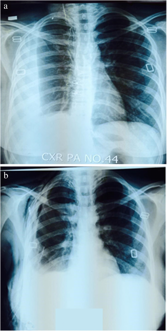
Loculated Pleural Effusion - Chest Radiograph Showing Right Loculated Pleural Effusion Download Scientific Diagram. Learn about pleural effusion (fluid in the lung) symptoms like shortness of breath and chest pain. Pleural effusion, also called water on the lung, is an excessive buildup of fluid between your lungs and chest cavity. Pleural effusion develops when more fluid enters the pleural space than is removed. A malignant pleural effusion may be large and diffuse or small and involve just a small portion of the pleural cavity. Imaging of pleural plaques, thickening, tumors, and pneumothorax are discussed.
Pleural fluid/serum ldh ratio >0.6. The imaging of pleural effusions will be presented here. Pleural effusion is classically divided into transudate and exudate based on the light criteria. Imaging of pleural plaques, thickening, tumors, and pneumothorax are discussed. Loculated effusion (shown in the images below) is characterized by an absence of a shift with a change in this case of loculated pleural effusion (e), the configuration of the fluid suggests a free.
Empyema Loculated Pleural Effusion Right Lateral Decubitus Radiograph Shows A Right Sided Pleural Effusion Whic Pleural Effusion Radiology Radiology Schools from i.pinimg.com Learn about different types of pleural effusions, including symptoms, causes, and treatments. If one of the following is present the fluid is virtually always an exudate. Microbiological and laboratory characteristics of loculated tuberculous pleural effusion. Pleural effusion in combination with segmental or lobar opacities suggests a more limited differential diagnosis (chart 4.3). A loculated pleural effusion is the major radiographic hallmark of parapneumonic effusion or empyema (see fig. The imaging of pleural effusions will be presented here. Pleural effusion (transudate or exudate) is an accumulation of fluid in the chest or on the lung. Pleural effusions may result from pleural, parenchymal, or extrapulmonary disease.
A joint effusion along with a pleural effusion may indicate an autoimmune disease.
Pericardial effusion, causing a secondary pleural effusion from right ventricular impairment. Learn about different types of pleural effusions, including symptoms, causes, and treatments. It can also be life threatening. Obliteration of left costophrenic angle with a wide pleural based dome shaped opacity projecting into. Pleural effusion with segmental and lobar opacities. Learn about pleural effusion including causes of pleural effusion. Case contributed by dr prashant mudgal. Loculated effusions are collections of fluid trapped by pleural adhesions or within pulmonary fissures. Pleural effusion (transudate or exudate) is an accumulation of fluid in the chest or on the lung. Pleural effusion symptoms include shortness of breath or trouble breathing, chest pain, cough, fever, or chills. A joint effusion along with a pleural effusion may indicate an autoimmune disease. Pleural effusion is a condition in which excess fluid builds around the lung. If none is present the fluid is virtually always a transudate.
A role in selected clinical circumstances. Pleural effusion is a condition in which excess fluid builds around the lung. Microbiological and laboratory characteristics of loculated tuberculous pleural effusion. A malignant pleural effusion can occur as a complication of cancer. Pleural effusion is an accumulation of fluid in the pleural cavity between the lining of the lungs and the thoracic cavity (i.e., the visceral and parietal pleurae).
The Loculated Pleural Effusion Revealed On Ct Scan Download Scientific Diagram from www.researchgate.net When a pleural effusion is loculated, the standard treatment methods of intercostal tube drainage and pleurodesis may not be helpful. Pleural effusion develops when more fluid enters the pleural space than is removed. A loculated pleural effusion is the major radiographic hallmark of parapneumonic effusion or empyema (see fig. Whereas, a heterogenous effusion with white septations indicates that it's loculated, and probably exudative. Case contributed by dr prashant mudgal. Learn about pleural effusion including causes of pleural effusion. Pleural effusions can loculate as a result of adhesions. Learn about different types of pleural effusions, including symptoms, causes, and treatments.
Pleural fluid/serum protein ratio >0.5.
Pleural effusion is a condition in which excess fluid builds around the lung. Whereas, a heterogenous effusion with white septations indicates that it's loculated, and probably exudative. Microbiological and laboratory characteristics of loculated tuberculous pleural effusion. A malignant pleural effusion may be large and diffuse or small and involve just a small portion of the pleural cavity. Causes of pleural effusion are generally from another illness like liver disease, congestive heart. A role in selected clinical circumstances. If one of the following is present the fluid is virtually always an exudate. Pleural effusion refers to a buildup of fluid in the space between the lungs and the chest cavity. It can also be life threatening. A malignant pleural effusion can occur as a complication of cancer. Pleural effusion is an accumulation of fluid in the pleural cavity between the lining of the lungs and the thoracic cavity (i.e., the visceral and parietal pleurae). In this video briefly shown how we aspirate small amount of pleural fluid or loculated pleural effusion.for more videos please subscribe the channel.if you. Case contributed by dr prashant mudgal.
Learn about pleural effusion including causes of pleural effusion. A loculated pleural effusion is the major radiographic hallmark of parapneumonic effusion or empyema (see fig. Pleural effusion with segmental and lobar opacities. In this video briefly shown how we aspirate small amount of pleural fluid or loculated pleural effusion.for more videos please subscribe the channel.if you. Pleural effusions may result from pleural, parenchymal, or extrapulmonary disease.
Role Of Medical Thoracoscopy In The Management Of Multiloculated Empyema Bmc Pulmonary Medicine Full Text from media.springernature.com Pleural fluid/serum ldh ratio >0.6. Pleural effusions can loculate as a result of adhesions. Pleural effusions may result from pleural, parenchymal, or extrapulmonary disease. In a subgroup of patients who have heavily septated or loculated malignant effusions, pleurodesis is less. Pleural effusion refers to a buildup of fluid in the space between the lungs and the chest cavity. If none is present the fluid is virtually always a transudate. Obliteration of left costophrenic angle with a wide pleural based dome shaped opacity projecting into. Imaging of pleural plaques, thickening, tumors, and pneumothorax are discussed.
Learn about pleural effusion including causes of pleural effusion.
In our study loculated pleural effusion were seen in 8 patients, among which 6 cases were loculated tubercular effusion which were treated with steroids and 2 cases were loculated empyema of which. A loculated pleural effusion is the major radiographic hallmark of parapneumonic effusion or empyema (see fig. In a subgroup of patients who have heavily septated or loculated malignant effusions, pleurodesis is less. Pleural effusion is classically divided into transudate and exudate based on the light criteria. Pleural effusions can loculate as a result of adhesions. Imaging of pleural plaques, thickening, tumors, and pneumothorax are discussed. If none is present the fluid is virtually always a transudate. A malignant pleural effusion can occur as a complication of cancer. Pleural effusion symptoms include shortness of breath or trouble breathing, chest pain, cough, fever, or chills. Pleural effusions accompany a wide variety of disorders of the lung, pleura, and systemic the presenting manifestations of pleural effusion are largely determined by the underlying disease. A malignant pleural effusion may be large and diffuse or small and involve just a small portion of the pleural cavity. Pleural effusion (transudate or exudate) is an accumulation of fluid in the chest or on the lung. Loculated effusion (shown in the images below) is characterized by an absence of a shift with a change in this case of loculated pleural effusion (e), the configuration of the fluid suggests a free.
Share :
Post a Comment
for "Loculated Pleural Effusion - Chest Radiograph Showing Right Loculated Pleural Effusion Download Scientific Diagram"


Post a Comment for "Loculated Pleural Effusion - Chest Radiograph Showing Right Loculated Pleural Effusion Download Scientific Diagram"